← All researchDiagnostics & reconstruction · 2026

Single-camera 3D PIV

3D velocity fields from one camera, using an engineered point-spread function and deep learning.
Under reviewarXiv preprint
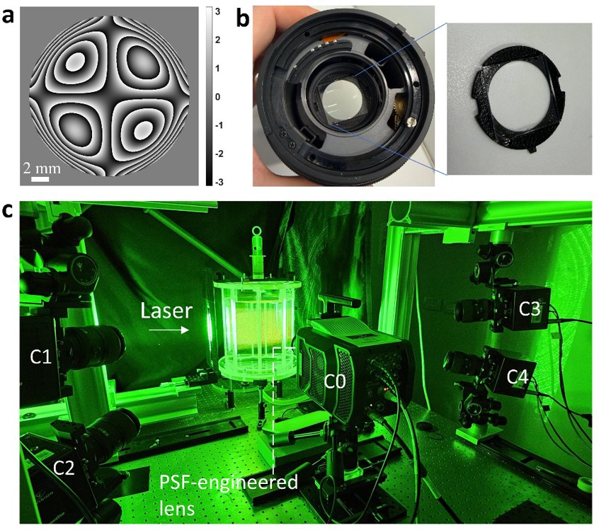
Phase mask, mask fitted in a macro lens, and the five-camera setup around a laser-illuminated tank
(a) Designed phase map of the mask in the PET camera (C0). (b) The mask inserted at the iris of a macro lens. (c) PET-PIV integrated into the tomographic PIV system with cameras C1–C4. From the arXiv preprint (2026), Fig. S2.

Overview

Most 3D PIV needs several cameras, and single-camera methods break down at high seeding density because particle images overlap. Many real experiments have room for only one camera.

Experimental approach

PET-PIV adds one thin phase mask to an otherwise standard PIV setup and calibrates the imaging system in place. Deep-learning algorithms then either track individual particles or, at very high density, reconstruct the 3D velocity field directly from short sequences of 2D images. I built the five-camera system that combines tomographic and PSF-engineered imaging, which gave the tomographic PIV reference for validation.

Key result

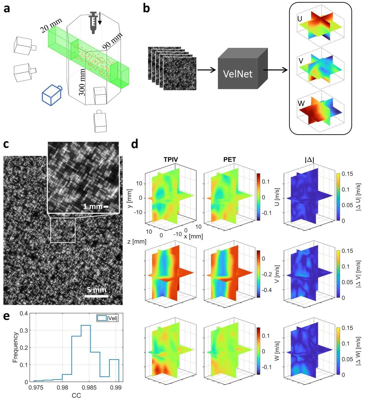
Against tomographic PIV, PET-PIV reached correlation coefficients above 0.97 and ran an order of magnitude faster, at seeding densities that previously needed several cameras.
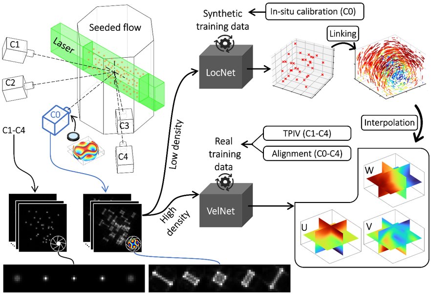
Schematic of PET-PIV with LocNet and VelNet processing paths
Schematic of PET-PIV. The camera C0 with a Tetrapod phase mask records depth-dependent particle images, while C1–C4 provide the tomographic reference. Sparse images are tracked with LocNet; dense images go to VelNet, which returns the 3D velocity field. From the arXiv preprint (2026), Fig. 1.
PET-PIV field mode compared with tomographic PIV
Field mode at macroscale: velocity slices from tomographic PIV and PET-PIV. From the arXiv preprint (2026).
← Back to researchBrowse publications →